3D Virtual Pancreatography
Shreeraj Jadhav, Konstantin Dmitriev, Joseph Marino, Matthew Barish, Arie Kaufman
External link (DOI)
View presentation:2021-10-27T14:00:00ZGMT-0600Change your timezone on the schedule page
2021-10-27T14:00:00Z
Fast forward
Direct link to video on YouTube: https://youtu.be/Pjrd657eGdY
Keywords
Visual diagnosis, Pancreatic cancer, Automatic segmentation, Lesion classification, Planar reformation, Pancreas, Three-dimensional displays, Ducts, Visualization, Computed tomography, Two dimensional displays
Abstract
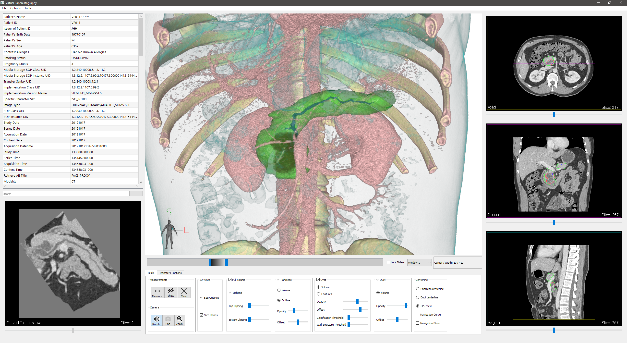
We present 3D virtual pancreatography (VP), a novel visualization procedure and application for non-invasive diagnosis and classification of pancreatic lesions, the precursors of pancreatic cancer. Currently, non-invasive screening of patients is performed through visual inspection of 2D axis-aligned CT images, though the relevant features are often not clearly visible nor automatically detected. VP is an end-to-end visual diagnosis system that includes: a machine learning based automatic segmentation of the pancreatic gland and the lesions, a semi-automatic approach to extract the primary pancreatic duct, a machine learning based automatic classification of lesions into four prominent types, and specialized 3D and 2D exploratory visualizations of the pancreas, lesions and surrounding anatomy. We combine volume rendering with pancreas- and lesion-centric visualizations and measurements for effective diagnosis. We designed VP through close collaboration and feedback from expert radiologists, and evaluated it on multiple real-world CT datasets with various pancreatic lesions and case studies examined by the expert radiologists.