Evaluation of cinematic volume rendering open-source and commercial solutions for the exploration of congenital heart data
Irum Baseer, Israel Valverde, Abdel H. Moustafa, Josep Blat, Oscar Camara
Room: 104
2023-10-25T03:45:00ZGMT-0600Change your timezone on the schedule page
2023-10-25T03:45:00Z
Fast forward
Full Video
Keywords
Cinematic rendering, open-source, commercial tool, congenital heart data
Abstract
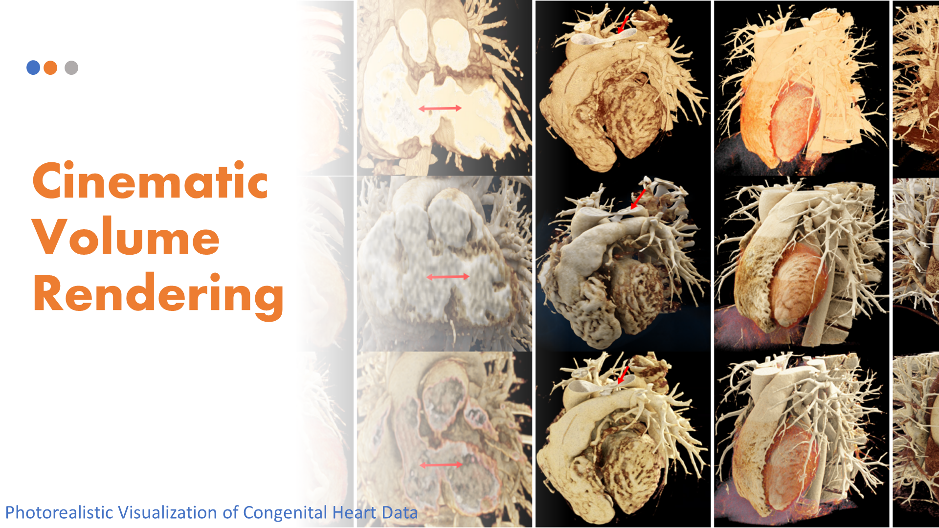
Detailed anatomical information is essential to optimize medical decisions for surgical and pre-operative planning in patients with congenital heart disease. The visualization techniques commonly used in clinical routine for the exploration of complex cardiac data are based on multi-planar reformations, maximum intensity projection, and volume rendering, which rely on basic lighting models prone to image distortion. On the other hand, cinematic rendering (CR), a three-dimensional visualization technique based on physically-based rendering methods, can create volumetric images with high fidelity. However, there are a lot of parameters involved in CR that affect the visualization results, thus being dependent on the user's experience and requiring detailed evaluation protocols to compare available solutions. In this study, we have analyzed the impact of the most relevant parameters in a CR pipeline developed in the open-source version of the MeVisLab framework for the visualization of the heart anatomy of three congenital patients and two adults from CT images. The resulting visualizations were compared to a commercial tool used in the clinics with a questionnaire filled in by clinical users, providing similar definitions of structures, depth perception, texture appearance, realism, and diagnostic ability.